TEST SITE
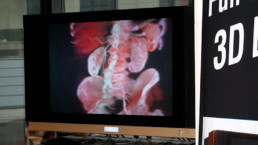
The use of state of the art cross sectional imaging significantly changed medical diagnostics. Cross-sectional imaging is usually used to refer to CT, MRI, PET, and SPECT and related imaging techniques that view the body in cross-section i.e. as axial (cross-sectional) slices.
Using these DICOM data we generate 3D volumetric data and if needed, the multiplanar (MPR) data visualization that are applied in numerous clinical fields. Today these images are visualized on conventional 2D monitors and 3D effects can only be achieved by “rotating” the viewed
image.
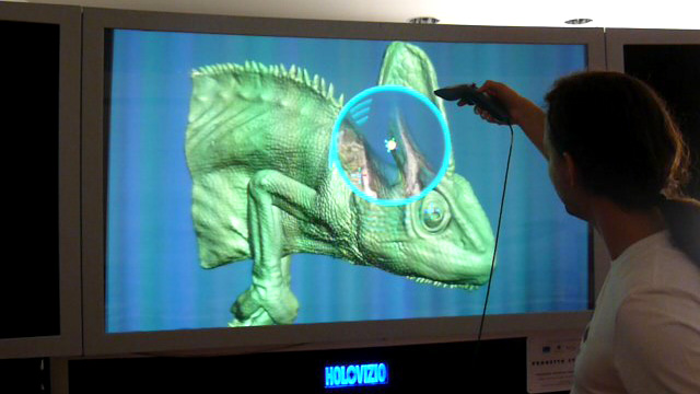
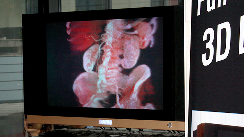
By the use of a secondary image processing of the visualized body or organ volume, 3D light field displaying offers an in depth examination option of organs like brain, spine or joints the vascular system. The normal anatomy or the pathological changes of the vessels can be recognized much easier using HoloVizio technology; the reconstructed image can be viewed without glasses by medical teams in a collaborative way, interaction is also possible, so this new technology can fundamentally change the interdisciplinary consultation of radiology examinations, surgical planning processes and medical education. It can be also applied in dental radiology.
If you have questions regarding compatibility, please check Software overview, contact us via contact form or write to
- 3D light field visualisation visible without glasses
- Extended, image-based diagnostic capabilities
- Ideal tool for surgical training and planning
- Decreased number of uncertainties and reduced examination time
